
Figure 1.

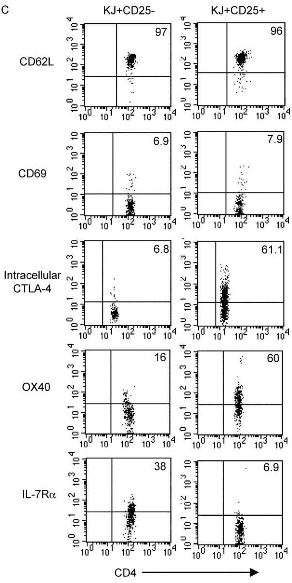
Abundant clonotype+ CD25+ cells in DO11 × RIP-mOVA double transgenic mice. (A) FACS® profiles of DO11 single positive versus DO11 × RIP-mOVA double transgenic littermates at 7 wk of age. Cells were isolated from the indicated lymphoid tissues and stained with CD4-PERCP, CD25-PE, and KJ-126-APC. Plots are gated on CD4+ KJ+ cells (except for thymus plots, which are gated on CD4+ CD8− KJ+ cells) and show the percentage of CD25+ cells. (B) Absolute number of CD4+ KJ+ CD25+ cells in the lymphoid tissues of DO11 single positive versus DO11 × RIP-mOVA double transgenic littermates. Data show mean and standard deviation from three mice of each genotype aged 6–7 wk. (C) Peripheral LN cells from DO11 × RIP-mOVA double transgenic mice were stained with various combinations of KJ-126-APC, CD62L-FITC, CD69PE, CD4-PERCP, OX40- biotin, streptavidin-PE, IL-7Rα-PE, and CD25-FITC/PE. For intracellular staining cells were surface stained and then permeabilized and stained with CTLA-4-PE. Expression profiles of each marker are shown for gated CD4+ KJ+ CD25− versus CD4+ KJ+ CD25+ cells.