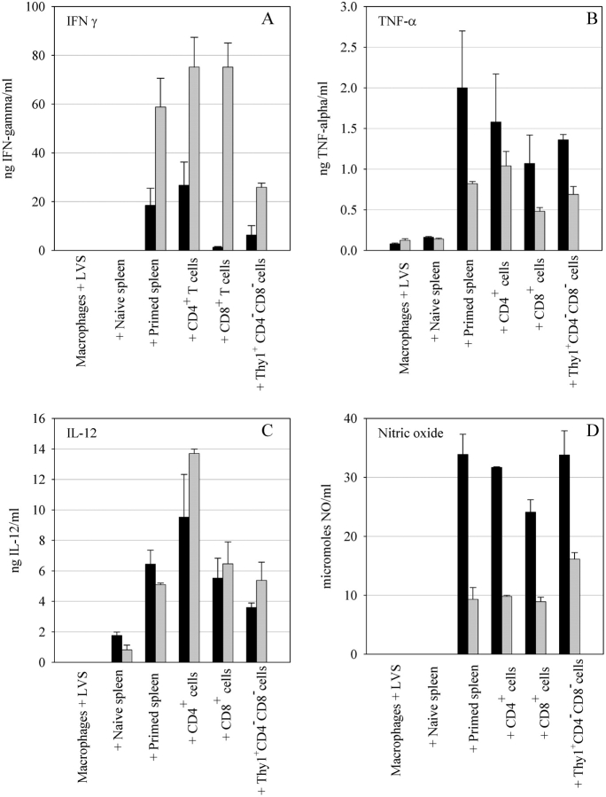
Figure 6.
Secretion of cytokines and NO into culture supernatants after coculture of LVS infected BMMØs and immune splenocytes. Wild-type (black bars) and IFNγR KO (gray bars) BMMØ were infected with LVS and cocultured with the indicated splenocyte populations. Splenocytes from either unprimed mice or mice infected 4 wk previously with an intradermal LVS infection were cocultured with LVS-infected macrophages at a 1:2 ratio (splenocytes to BMMØ). Similarly, CD4+ and CD8+ and Thy1+CD4−CD8− splenocyte subpopulations were added to infected BMMØ cultures at a 1:2 ratio. Culture supernatants were collected 72 h later and tested for IFN-γ (A), TNF-α (B), IL-12 (C), and NO (D). Values shown are the mean ng/ml ± the SEM of the indicated cytokine (A–C) or mean μM/ml nitrite (D) (triplicate samples). These results are representative of three experiments of similar design.

