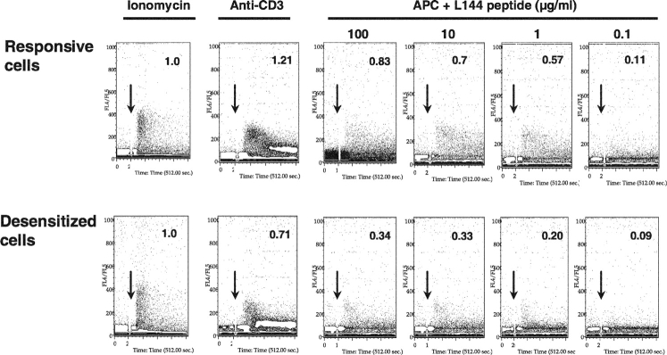
Figure 7.
Impairment in sustained calcium influx in the desensitized cells. T cells of the responsive and desensitized cell lines were loaded with the indicator dye Indo-1-AM. Calcium mobilization of the cells was monitored continuously by flow cytometry upon stimulation of the T cells with ionomycin, cross-linking anti-CD3, or with I-As/B7 expressing DAS cells, that had been loaded with the indicated concentrations of L144 peptide (start of stimulation indicated by arrow). Calcium mobilization in antibody and antigen stimulated cells was measured and compared with activation with ionomycin, by comparing the number of gated events above a baseline in the total time after stimulation (converted to arbitrary units and taking flux induced with ionomycin as 1.0).

