
Figure 7.
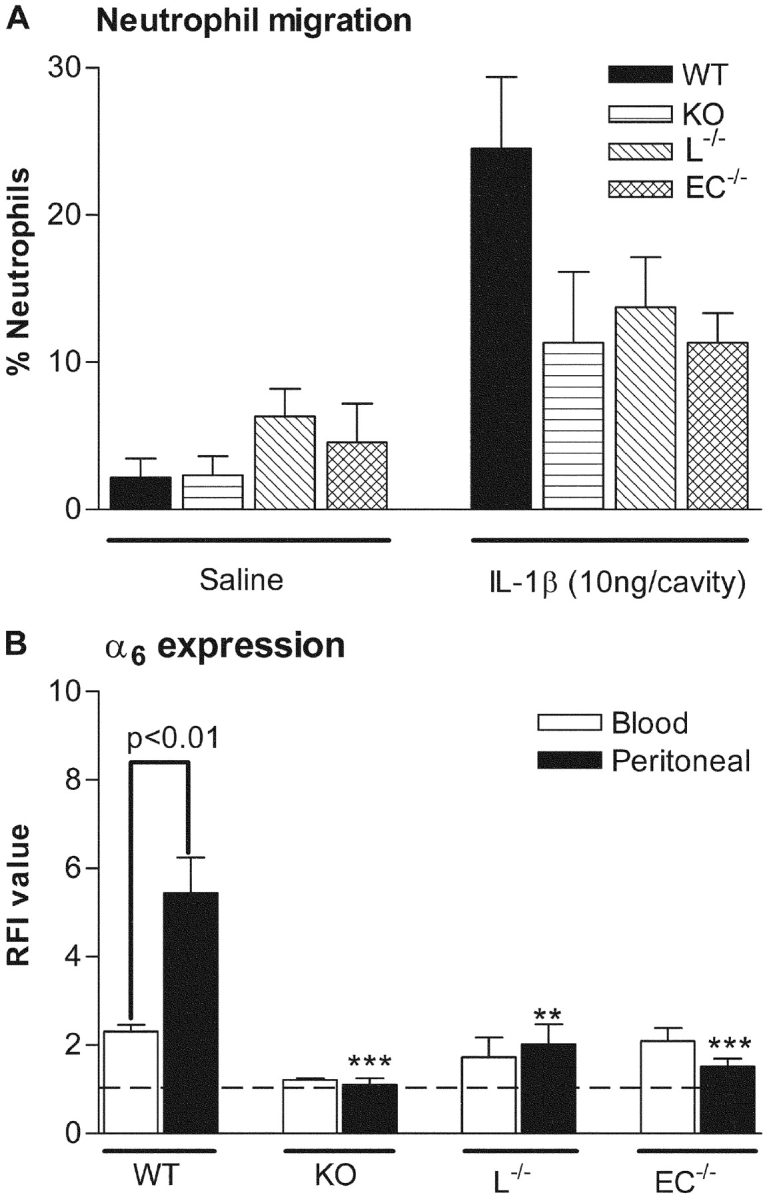
Neutrophil migration and cell surface expression of α6 on blood and peritoneal neutrophils in an IL-1β–elicited peritonitis model in WT, PECAM-1–deficient (KO), and chimeric mice deficient in either leukocyte (L−/−) or endothelial cell (E−/−) PECAM-1. Mice were injected via the intraperitoneal route with IL-1β (10 ng/animal) and 4 h later, blood or peritoneal lavage was collected. The lavage fluid was used to quantify neutrophil infiltration (A). Lavage and blood samples were also analyzed for the binding of GoH3 (anti-α6) as quantified by FACS® (B), as detailed in Materials and Methods. The data represent mean ± SEM of samples obtained from n = 4–7 animals/group. Significant differences between peritoneal samples from WT mice and KO, L−/−, or E−/− mice are indicated by **P < 0.01 and ***P < 0.001. Other statistical differences are indicated by lines.