
Figure 5.
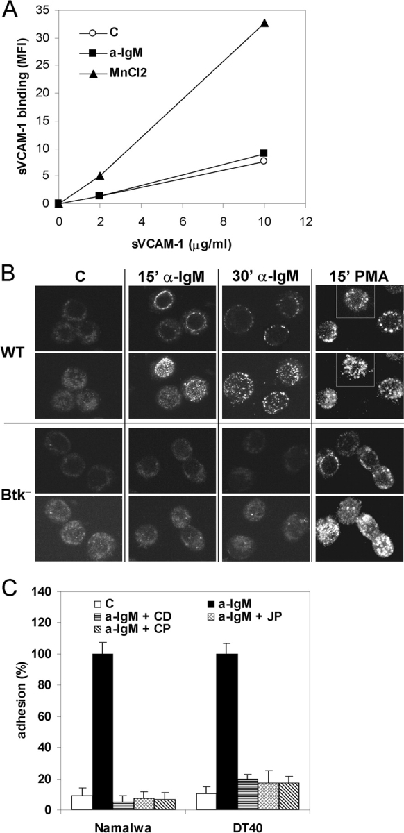
BCR-controlled integrin α4β1-mediated adhesion involves integrin clustering and cytoskeletal reorganization. (A) DT40 cells were incubated without (open circles) or with 10 μg/ml anti-IgM (closed squares) or 3 mM MnCl2 (closed triangles) and 0, 2, or 10 μg/ml soluble VCAM-1-Fc, as indicated. Binding of soluble VCAM-1-Fc was measured by FACS analysis. Mean fluorescence intensity (MFI) values of a representative experiment are depicted. (B) DT40 cells, either WT or Btk-deficient (Btk−), were not stimulated (C) or stimulated for 15 or 30 min with anti-IgM (a-IgM) or PMA, as indicated. Clustering of integrin α4β1 was analyzed by means of confocal microscopy. For each condition, representative images of optical midheight sections (1st and 3rd row), and corresponding whole cell projections along the z-axis using maximum fluorescence values (2nd and 4th row), are shown. (C) Namalwa cells (left) or DT40 cells (right) were pretreated for 30 min with 1 μg/ml Cytochalasin D (CD), 1 μM Jasplakinolide (JP), 100 μg/ml Calpeptin (CP), or left untreated, and subsequently not stimulated (C) or stimulated with anti-IgM (a-IgM) and plated on a surface coated with VCAM-1.