Figure 3.
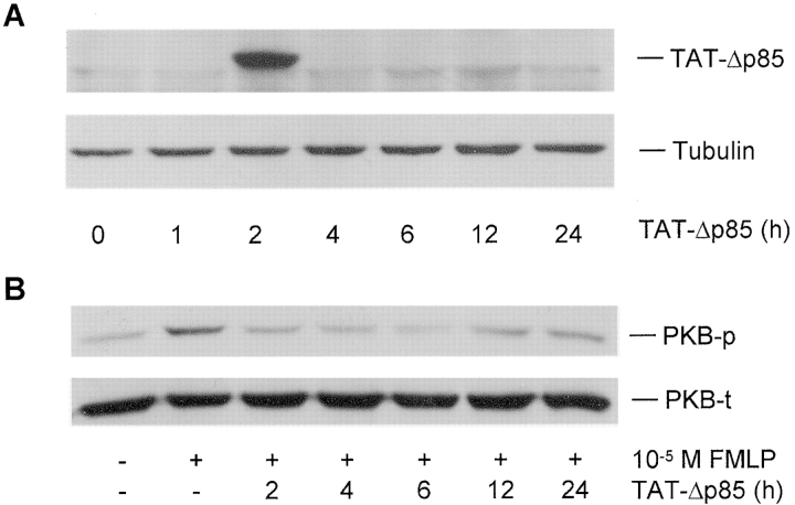
Western blot analysis of the uptake of TAT-Δp85 into lung tissue and the efficacy of TAT-Δp85 protein transduction on FMLP-induced PKB phosphorylation in lung tissues. (A) Transduction of TAT-Δp85 into the lung. Lungs were excised from mice at indicated times after i.p. administration of TAT-Δp85 (10 mg/kg), and lung tissue extracts were mixed with loading buffer and separated by SDS-PAGE, and probed by anti-His antibody. (B) Western blot analysis of the effect of TAT-Δp85 on FMLP-induced PKB phosphorylation in whole-lung tissue. FMLP was instilled intranasally at 0, 2, 4, 6, 12, and 24 h after administration of TAT-Δp85. Whole-lung cell extracts were prepared and analyzed by Western blot with antiphosphorylation-specific PKB Ab (top). Equivalency of loading was established for each lane with anti-PKB Ab, which measures total PKB (phosphorylated and nonphosphorylated) (bottom). Inhibition of PKB phosphorylation is maximal at 2–6 h, corresponding to fluorescence data from Fig. 2. By 12 h, there is diminished inhibition of PKB phosphorylation by TAT-Δp85. The result shown is representative of three different experiments.

