
Figure 1.
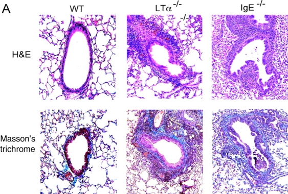


Increased inflammation and airway remodeling in LTα−/− and IgE−/− mice. (A) Lung tissues from WT, LTα−/−, and IgE−/− mice (3 mo old) were fixed in 10% buffered formalin and embedded in paraffin. Hematoxylin and eosin staining (top, H&E) and Masson's trichrome staining (bottom) of B6, LTα−/−, and IgE−/− mice are presented. (B) The lung (left) and BAL cells (right) were isolated by collagenase digestion and lavage, respectively, from 3-mo-old B6 (white bars) and LTα−/− (black bars) mice (n = 5 per group). The total number of cells was analyzed with trypan blue staining. The number of leukocytes in the lung of LTα−/− mice is significantly higher than that of the WT (P < 0.001). BAL cells from five mice were pooled for further analysis by FACS®. (C) Lung cells were dually stained with FITC-conjugated anti-CD69 and PE-conjugated anti-CD3. The fluorescence intensity was analyzed from the gated lymphocyte population of B6 (left) and LTα−/− (right) mice. The percentage of CD69+ cells among CD3+ cells is presented. (D) The forward/side scatter dot-plot profiles were analyzed from BAL cells of B6 (left) and LTα−/− (right) mice; (a), (b), and (c) represent the lymphocyte, granulocyte, and macrophage population, respectively. (E) The gated lymphocyte population (a) from the forward/side scatter dot-plot profiles of BAL cells (D) was analyzed for CD4 and B220 expression by FACS® analysis. (F) CCR3 and CD3 expression was analyzed from both the gated lymphocyte (a) and granulocyte (b) populations. (G) The type of BAL cells was determined by calculating the absolute number of each cell type from the FACS® profiles and by the total number of cells (P < 0.0001; WT vs. LTα−/−).
(H) B6 and LTα−/− mice were analyzed by three-color staining with FITC–anti-B220, cychrome–anti-CD4, and PE–anti-CD8 or PE–anti-NK1.1. The gated lymphocyte population (a) from the forward/side scatter dot-plot profiles of the lung cells was analyzed for CD4 and B220 (P < 0.01, WT vs. LTα−/− mice). The percentage of CD8+ or NK1.1+ cells gated from B220−CD4− was evaluated. (I) CCR3 and CD3 expression was analyzed from both the gated lymphocyte (a) and granulocyte (b) population. (J) The total number of cells was calculated. The type of lung cells was also determined by calculating the absolute number of each cell type from the FACS® profiles. The results are representative of five independent experiments.