
Figure 3.
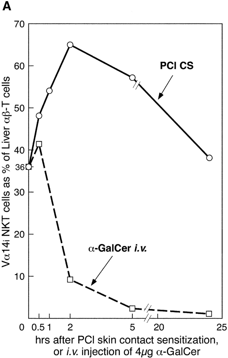

Liver NKT cells are activated early after PCl skin sensitization. (A) CBA/J mice were PCl skin sensitized or injected i.v. with 4 μg α-GalCer. Separate groups of mice were killed at 30 min, 1 h, 2 h, 5 h, or 24 h, and LMNCs were isolated and stained with FITC anti-TCRβ mAb and PE CD1d–α-GalCer tetramers and analyzed by flow cytometry to identify double positive Vα14i NKT cells. Percentages were determined based on the analysis of pooled LMNCs from four to five mice per group, except for 2- and 5-h points performed twice. (B) Dot plots show the percentage of tetramer-positive T cells from a representative experiment comparing nonimmune and 2-h skin immune CBA/J mice.