
Figure 3.
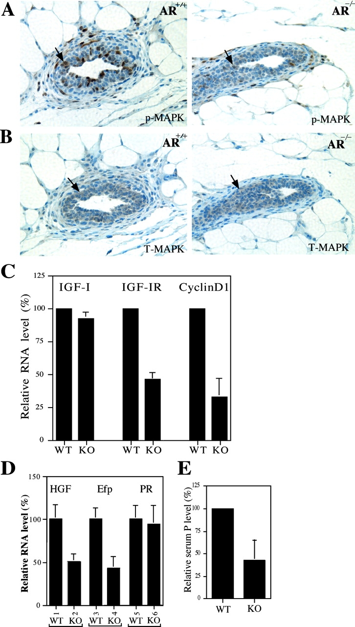
The reduced MAPK activity and mRNA expression of IGF-IR, HGF, and Efp in AR −/− mammary glands. (A) Reduced MAPK activities are observed in AR −/− mammary glands of 4-wk-old mice. Mammary glands were stained with anti–phospho-MAPK (p-MAPK) antibody. The decrease of positive staining (brown) indicates the reduced MAPK activity in AR −/− mammary glands. (B) The similar total MAPK (T-MAPK) staining results are observed in the mammary glands from 4-wk-old AR +/+ and AR −/− mice. Adjacent sections were used for p-MAPK and T-MAPK stainings. (C) The mRNA expression of IGF-IR, but not IGF-I, is reduced in AR −/− mice. Total RNA was extracted from 4-wk-old AR +/+ and AR −/− mice and quantitated by real-time RT-PCR. Cyclin D1, a proliferation indicator, is also reduced in mammary glands of female AR −/− mice. (D) The mRNA expressions of two ER target genes, HGF and Efp, are reduced in AR −/− mice. The expression of PR mRNA has no significant difference (bars 5 vs. 6). Total RNA was extracted from 4-wk-old AR +/+ and AR −/− mice injected with E2 (n = 5 for each group). (E) The serum levels of P were reduced in 12–16-wk-old adult female AR −/− mice.