
Figure 7.
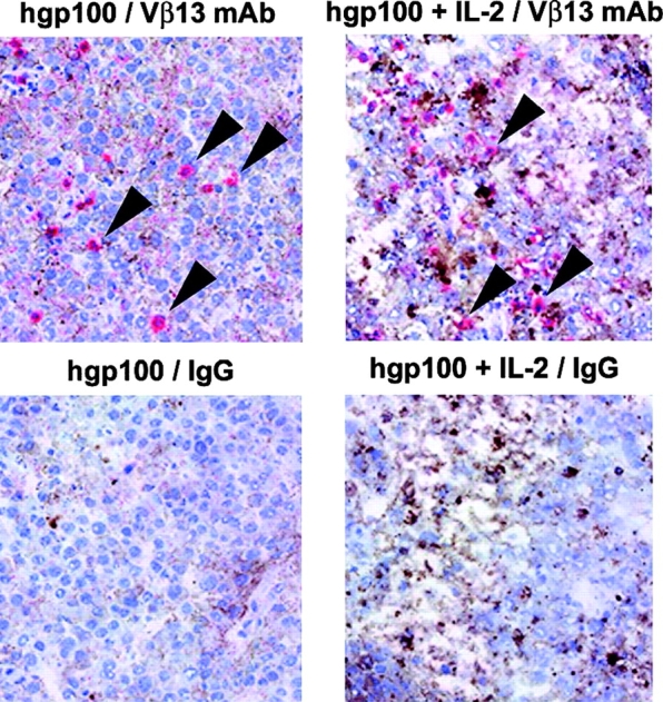
Histological analysis reveals presence of Vβ13+ T cells in tumors and demonstrates the activating effects of IL-2. T cells and tissue architecture within tumors were visualized after vaccination with or without IL-2. C57BL/6 mice bearing 14-d B16 tumors received 107 pmel-1 splenocytes and vaccination with hgp10025–33 peptide in IFA and anti-CD40 with or without IL-2. As was the case for vaccination with rVVs or rFPVs encoding hgp100, vaccination with hgp10025–33 peptide in IFA and anti-CD40 after the adoptive transfer of pmel-1 cells was greatly enhanced with the addition of IL-2 in repeated experiments (not depicted). Tumors were excised 7 d after the vaccination and 4-μm frozen sections were stained with anti-Vβ13 mAb (top, arrows) or IgG control (bottom), counterstained with hematoxylin, and photographed at an original magnification of 200. (left) pmel-1 cells plus hgp100 vaccination. (right) pmel-1 cells plus hgp100 vaccination followed by IL-2 administration.