
Figure 5.
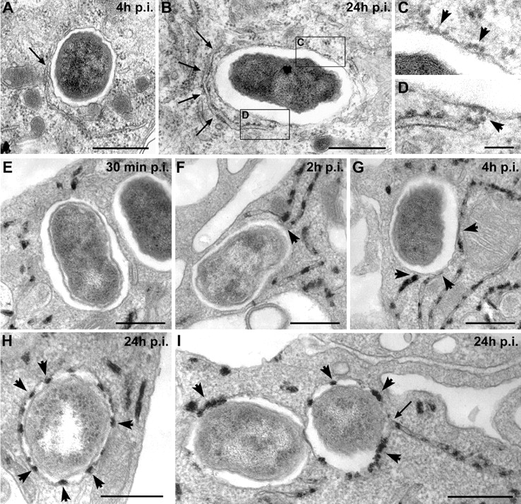
BCVs acquire ER membranes through limited fusion with the ER. BMDM were infected with the Brucella wild-type strain 2308 for various times. Samples were processed for conventional EM (A–D) or EM cytochemistry for G6Pase detection (E–I). (A) BCVs at 4 h after infection showing a close contact with the ER (arrow). (B) BCVs at 24 h after infection surrounded by ER (arrows). (C) Enlarged view of the vacuolar membrane from B showing ribosomes studding the membrane (arrowheads). (D) Enlarged view of the contact site between ER and the vacuolar membrane showing ribosomes on the vacuolar membrane (arrowhead). (E) BCVs at 30 min after infection showing no interaction with G6Pase+ ER. (F) BCVs at 2 h after infection in close contact with G6Pase+ ER (arrowheads). (G) BCVs at 4 h after infection in intimate contact with several G6Pase+ ER compartments (arrowheads). (H) Representative G6Pase+ late BCVs at 24 h after infection (arrowheads show the intravacuolar G6Pase reaction product). (I) G6Pase+ late BCVs at 24 h after infection. The right-hand side BCV is fusing with the ER (arrow). Bars, 1 μm (A and B), 0.2 μm (C and D), and 0.5 μm (E–I).