Figure 8.
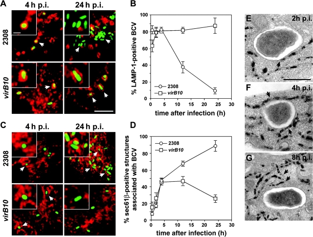
VirB mutant–containing vacuoles fail to sustain fusion-proficient interactions with the ER. BMDM were infected with either the wild-type Brucella strain GFP-2308 or the mutant strain GFP-virB10 for various times. Samples were processed for immunofluorescence (A–D) or EM staining for G6Pase (E–G). (A) Confocal images of infected BMDM showing acquisition of LAMP-1 on 2308 or virB10-containing vacuoles after 4 or 24 h of infection. (B) Quantitation of LAMP-1 acquisition by 2308 (○)- or virB10 (□)-containing vacuoles. Data are means ± SD of three independent experiments. (C) Confocal images of GFP Brucella–infected BMDM showing association with, or acquisition of, Sec61β+ structures on 2308 or virB10-containing vacuoles after 4 or 24 h of infection. (D) Quantitation of association of Sec61β1 structures with 2308 (○)- or virB10 (□)-containing vacuoles. Data are means ± SD of five independent experiments. (E) Staining for G6Pase in virB10-infected BMDM at 2 h after infection. The arrowhead shows a close association of BCVs with ER. (F) Staining for G6Pase in virB10-infected BMDM at 4 h after infection. The BCV is surrounded by ER (arrowheads). (G) Staining for G6Pase in virB10-infected BMDM at 8 h after infection Arrowheads show that the ER is no longer in the close vicinity of the BCV. G6Pase reaction product is not detectable inside the BCV. Bars, 5 μm (A and C), 1 μm (insets in A and C), and 0.5 μm (E–G).

