
Figure 6.
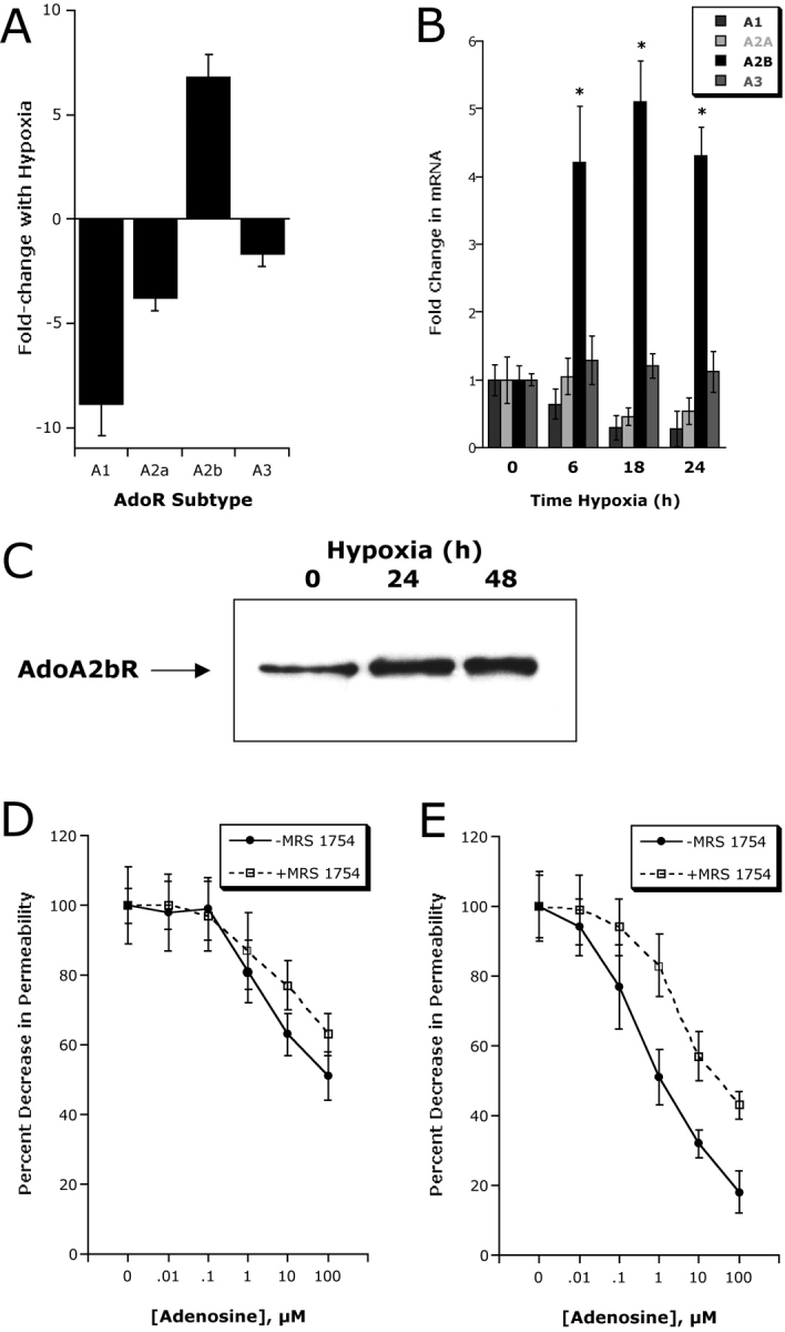
Adenosine A2B receptor (AdoRA2B) induction by hypoxia enhances barrier response. (A) Microarray analysis of individual adenosine receptors in response to hypoxia (AdoRA1, AdoRA2A, AdoRA2B, and AdoRA3). Confluent HMEC-1 were exposed to normoxia or hypoxia (12 h exposure to pO2 20 torr) and the relative expression of individual adenosine receptors was quantified from total RNA by microarray analysis. Data are expressed as fold change ± SD relative to normoxia. (B) Real-time PCR analysis was employed to confirm hypoxia-regulated expression of individual adenosine receptors (AdoRA1, AdoRA2A, AdoRA2B, and AdoRA3) in HMEC-1. Data were calculated relative to internal control (β-actin) and are expressed as fold increase ± SD over normoxia at indicated time points. Results are derived from three experiments in each condition (*, P < 0.01 compared with normoxia). (C) Increased AdoRA2B surface protein with hypoxia. Confluent HMEC-1 monolayers were exposed to indicated periods of hypoxia, monolayers were washed, surface proteins were biotinylated, and cells were lysed. AdoRA2B was immunoprecipitated with mAb to human AdoRA2B and resolved by SDS-PAGE, and resultant Western blots were probed with avidin-peroxidase. A representative experiment of three is shown. (D) Hypoxia induction of AdoRA2B enhances barrier response of HMEC-1 to adenosine. Indicated concentrations of adenosine were added to HMEC-1 monolayers preexposed to normoxia or hypoxia (48 h). Addition of the specific AdoRA2B-antagonist MRS 1754 (100 nM) significantly shifted the adenosine dose response in posthypoxic endothelium (P < 0.01 by ANOVA). Data are derived from 6 monolayers in each condition. Data are expressed as mean ± SD of percent control flux.