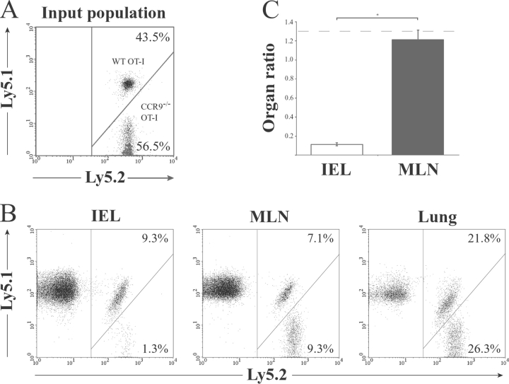
Figure 4.
OT-1 lymphocyte localization to the small intestinal epithelium after immunization with OVA and adjuvant is CCR9 dependent. CCR9−/− (Ly5.1−Ly5.2+) and WT (Ly5.1+Ly5.2+) OT-1 cells were coinjected (3–5 × 10⁶ total cells) i.v. into C57BL/6J-Ly5.1 (Ly5.1+Ly5.2−) mice. Mice received OVA and LPS i.p. 2 d after cell transfer, and the percentage of CCR9−/− and WT OT-1 cells among CD8β1T cells was determined in each organ by flow cytometry analysis 3 d later. (A and B) Representative flow cytometry analysis from one experiment of three performed. (C). The CCR9−/− to WT OT-1 cell ratio in the MLN and IEL. Organ ratio was determined by dividing the percentage of CCR9−/− (Ly5.2+) OT-1 cells with the percentage of WT (Ly5.1+Ly5.2+) OT-1 cells in each organ. Results are mean (SEM) of four mice in each group and from one representative experiment of three performed. *P = 0.0286. Dotted line represents the CCR9−/− to WT OT-1 cell input ratio.

