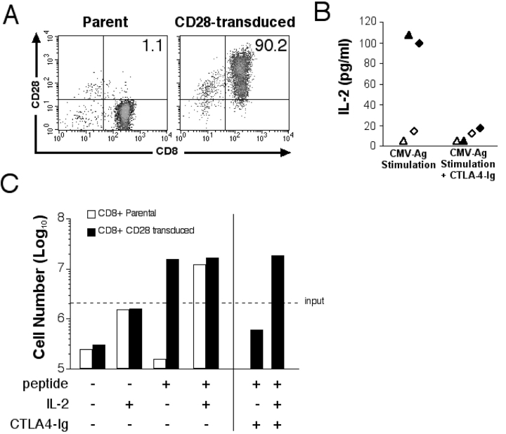
Figure 3.
Reconstitution of CD28 restores Ag-triggered IL-2 production and T cell proliferation. (A) CD28 expression was assessed before and after transduction of a CMV-specific CD28− CD8+ T cell clone, MT-2 with the CD28 gene. The percentage of cells positive for both CD8 and CD28 is indicated and is representative of five experiments. (B) IL-2 production by parental and CD28-transduced CMV-specific CD8+ T cells after Ag stimulation. HLA-matched peptide-pulsed LCLs (CD80/86+) were incubated with parental (⋄ and ▵) or CD28-transduced (♦ and ▴) CMV-specific CD8+ T cell clones MT-2 (▴ and ▵) and MT-3 (♦ and ⋄). CTLA-4–Ig was added to indicated wells. Data points represent the means of two samples for each clone. (C) Proliferation of CD28− parental and CD28-transduced CD8+ CMV-specific T cells. 2 × 10⁶ parental (white bar) and CD28-transduced (black bar) T cells were plated with 2 × 10⁶ mitomycin C–treated T2 cells alone or pulsed with 100 nM of pp65495–503 peptide. IL-2 (10 U/ml) and CTLA-4–Ig were added to selected wells. Cell growth was assessed at day 5 by counting viable T cells.

