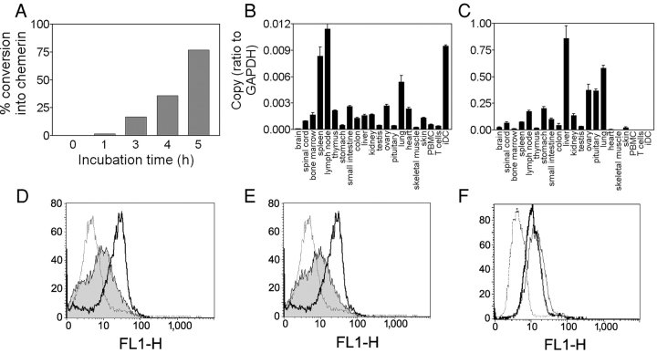
Figure 4.
Expression of human chemerin and its receptor. (A) Conversion of 100 nM human recombinant prochemerin in conditioned medium from hamster CHO-K1 cells. Conversion rate was estimated by comparing the biological activity with that of the same molar amount of purified processed chemerin. (B and C) Transcripts encoding human chemerinR (B) and prochemerin (C) were amplified by quantitative RT-PCR in a set of human tissues and cell populations. PBMC, peripheral blood mononuclear cells; iDC, immature DCs. (D and E) The expression of chemerinR was analyzed by FACS® in immature (solid line) and mature DCs (gray area) after stimulation by LPS (D) or CD40L (E), using the 1H2 monoclonal antibody (IgG2A). Control labeling (dotted line) was made with an antibody of the same isotype. (F) ChemerinR expression on macrophages was monitored using the 1H2 (thick solid line) and 4C7 (thin solid line) monoclonal antibodies. Control labeling (dotted line) was made with an antibody of the same isotype.

