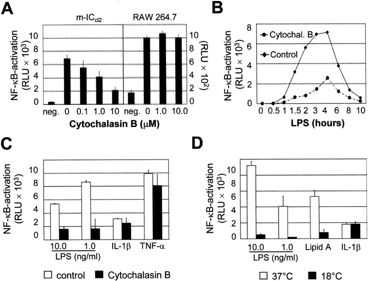
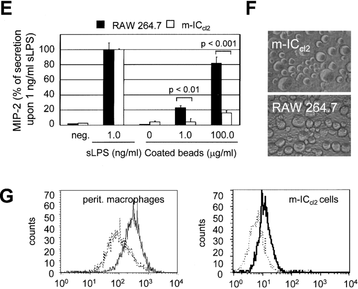
Figure 1.
Requirement of intact cell traffic for LPS-mediated cell activation. (A) Effect of cytochalasin B on LPS-induced cellular stimulation of m-ICcl2 cells and RAW 264.7 cells carrying an NF-κB luciferase reporter construct. Data are presented as relative light units (RLU). (B) Time course of luciferase production after LPS stimulation (10 ng/ml) in the presence or absence of cytochalasin B. (C) Comparison of the inhibitory effect of 10 μM cytochalasin B on cellular stimulation mediated by LPS, lipid A, IL-1β, or TNF-α. (D) Effect of the incubation temperature on LPS-mediated NF-κB stimulation. Stimulation was performed with 10.0 or 1.0 ng/ml LPS, 10 ng/ml lipid A, or 100 ng/ml IL-1β at 37 or 18°C for 2 or 6 h, respectively. (E) Comparison of MIP-2 secretion by macrophage-like RAW 264.7 cells and intestinal epithelial m-ICcl2 cells in response to soluble LPS (sLPS) versus LPS covalently linked to agarose beads. The data are presented as percent of MIP-2 secretion obtained after exposure to 1.0 ng/ml sLPS. Agarose beads were coated in the absence or presence of 1.0 or 100.0 μg/ml LPS. (F) The micrograph illustrates the situation of RAW 264.7 and m-ICcl2 cells exposed to agarose beads. ×200. (G) FACS® analysis for TLR4 on primary macrophages and m-ICcl2 cells using the rat monoclonal anti–TLR4/MD-2 antibody MTS510. Dotted line, isotype control.


