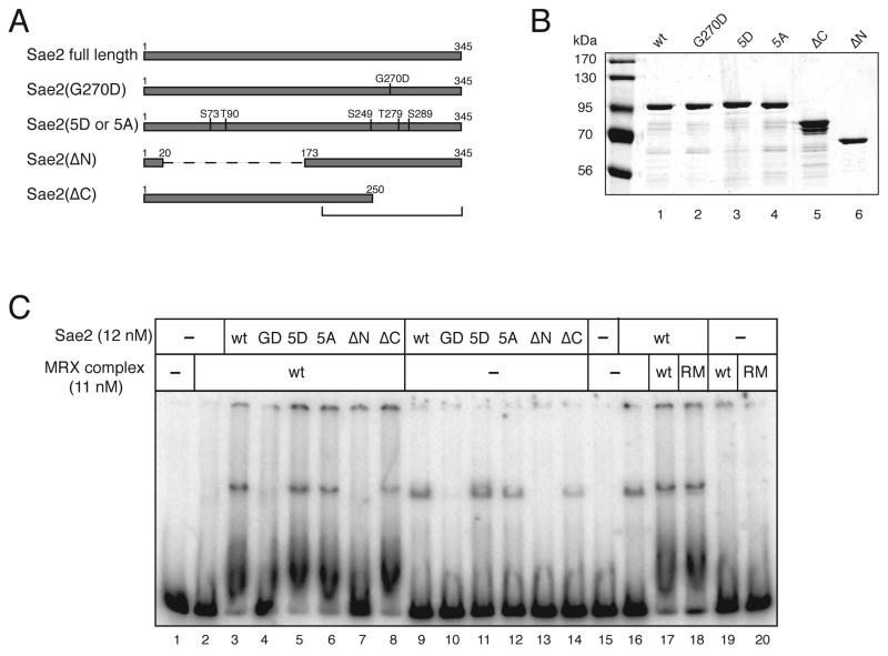
Figure 1.
Expression and DNA binding activity of recombinant Sae2. (A) Schematic representation of full-length wild-type Sae2, and mutants G270D, 5D (residues indicated changed to aspartate), 5A (residues indicated changed to alanine), ΔN (Δ a.a. 21 to 173) and ΔC (Δ a.a. 251 to 345). (B) SDS-PAGE of purified Sae2 wild-type and mutant proteins, approximately 100 ng total protein each. (C) Wild-type (wt) and mutant Sae2 proteins were incubated with a 249 bp double-stranded DNA substrate and analyzed in a 8% native polyacrylamide gel in the presence of wild-type (wt) or R20M (RM) MRX complex. Protein-DNA complex 1 and complex 2 are indicated (described in text).

