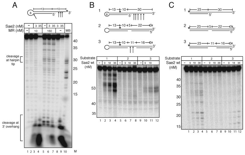
Figure 3.
Sae2 cleaves hairpin DNA. (A) Wild-type MR and Sae2 were incubated with a 3′ [32P]-labeled hairpin DNA substrate with 1 mM MnCl2 and 0.5 mM ATP as indicated and separated in a denaturing polyacrylamide gel. Substrate was also incubated with Mung Bean nuclease as a control to show the location of hairpin cut at the tip (MB). Numbers in the “M” lane indicate the positions of DNA standards run in the same gel. (B) Reactions were performed as in (A) with Sae2 only, 5 mM MgCl2, and internally-labeled hairpin substrates as shown. (C) Reactions were performed with substrates identical to those in (B) except lacking the hairpin loops.

