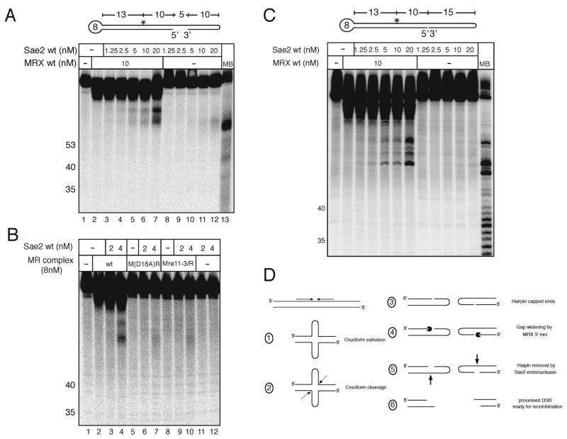
Figure 5.
Mre11 exonuclease activity facilitates Sae2 hairpin removal. (A) An internally [32P]-labeled double hairpin DNA substrate was incubated first with wild-type MRX in 1 mM MnCl2 for 20 minutes; Sae2 was then added with 5 mM MgCl2 and the reactions were continued for an additional 30 minutes before separation on a denaturing polyacrylamide gel. MB control as in Fig. 3. (B) Reactions were performed as in (A) but with wild-type MR and Mre11 nuclease-deficient complexes M(D16A)R and M(Mre11-3)R as indicated. (C) Hairpin cleavage assays as in (A) except that the substrate has a nick instead of a gap. (D) Model of MRX/Sae2 hairpin processing. Dotted arrow represents predicted MRX/Sae2-independent cleavage of the cruciform; Pacman represents MRX 3′ to 5′ exonuclease activity; bold arrow represents Sae2 cleavage. The location of the cruciform cleavage site is assumed to be at the base of the cruciform as shown but this has not been demonstrated in vivo.

