Figure 6.
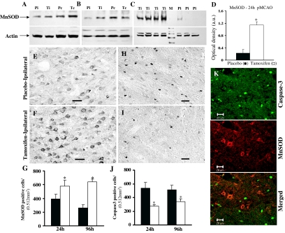
Effect of tamoxifen on MnSOD expression and caspase-3 activation in the ischemic cortex after pMCAO. A and B, Western blot analysis reveals that MnSOD protein levels are greatly reduced in the ischemic cortex (Pi) compared with contralateral cortex (Pc) of placebo-treated or sham-operated animals (not shown) after 1 h (A) and 4 h (B) pMCAO. This decrease in MnSOD expression was largely prevented in the ischemic cortex of tamoxifen-treated (Ti) animals (n = 4–5 per group). B, A similar reduction in the MnSOD protein levels was observed in the ischemic cortex of placebo-treated (Pi, represents three individual animals) but not in tamoxifen-treated (Ti, represents four individual animals) animals after 24 h pMCAO. D, Statistical bar diagram of MnSOD protein levels (as shown in panel C) after correction with the housekeeping protein β-actin after 24 h pMCAO. E and F, MnSOD-DAB staining performed on coronal sections confirmed a significant reduction in MnSOD-immunoreactive cells in the ischemic cortex of placebo-treated (E) compared with tamoxifen-treated (F) animals after 24 pMCAO (n = 4–5 per group). G, Statistical analysis of MnSOD-positive cells performed after 24 and 96 h pMCAO shows a significant (P < 0.05) reduction in placebo-treated (black bars) compared with tamoxifen-treated (white bars) animals. H and I, Caspase-3-DAB staining performed on coronal sections showed a significantly higher number of active caspase-3-immunoreactive cells in the ischemic penumbra of placebo-treated animals (H) compared with tamoxifen-treated animals (I) at 24 h pMCAO (n = 4–5 per group). J, Statistical analysis of caspase-3-positive cells performed after 24 and 96 h pMCAO shows a significant reduction in placebo (black bars) compared with tamoxifen-treated (white bars) animals. *, Significance was determined by one-way ANOVA with post hoc analysis using Student-Newman-Keuls test; P < 0.05. K, Confocal laser microscopy performed on coronal sections double-labeled for active caspase-3 (green) and MnSOD (red) indicate that these two proteins do not colocalize. Scale bars, 20 μm.

