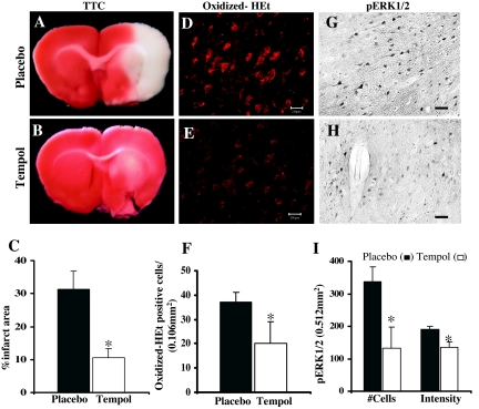
Figure 7.
Effect of tempol on infarct volume, O2− production, and ERK1/2 activation after pMCAO. Compared with placebo-treated animals (A and C), tempol-treated animals (B and C) show a significantly reduced infarct area at 24 h pMCAO. D–F, O2− production, as measured by oxidized-HET signal, was significantly reduced (P < 0.05) in ischemic penumbra cortex of tempol-treated animals (E and F) compared with placebo-treated animals (D and F) after 2 h pMCAO (n = 6 in each group). Tempol treatment (H and I) also markedly reduced ERK1/2 activation in the penumbra compared with placebo-treated animals (G and I) at 2 h after pMCAO. *, Significance was determined by one-way ANOVA with post hoc analysis using Student-Newman-Keuls test; P < 0.05. Scale bars, 20 μm.

