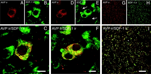
Figure 1.
Confocal microscopic images of sections from the PVN (A–C), SON (D–F), and posterior pituitary (G–I) of control (LE) rats immunolabeled with SDF-1 (green) and vasopressin (red). Dually labeled neurons stand out in yellow when images are merged. *, Blood vessels; ←, dendrite processes. Scale bars, 10 μm (PVN and SON); 50 μm (posterior pituitary).

