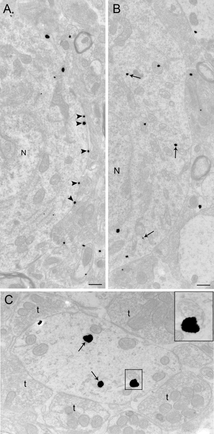
Figure 3.
Electron microscopic evidence for stress-induced CRFr internalization in LC perikarya and dendrites. A and B, Electron photomicrographs showing CRFr cellular localization in perikarya from a control rat (A) and from a rat perfused 24 h after 15-min swim stress (B). A, Immunogold-silver labeling for CRFr (arrowheads) along the plasma membrane in a control case; B, CRFr labeling shifts from the plasma membrane to the cytoplasm 24 h after 15-min swim stress. Arrows point to immunogold-silver labeling in the cytoplasm, whereas arrowheads point to immunogold-silver labeling on the plasma membrane. N, Nucleus. C, Dendrite from a 24-h post-stress subject showing CRFr in the cytoplasm and association with a multivesicular body. The dendrite is targeted by multiple axon terminals (t). The inset shows a higher-magnification view of the area denoted by the box in C where CRFr is associated with a multivesicular body. Scale bar, 0.5 μm.

