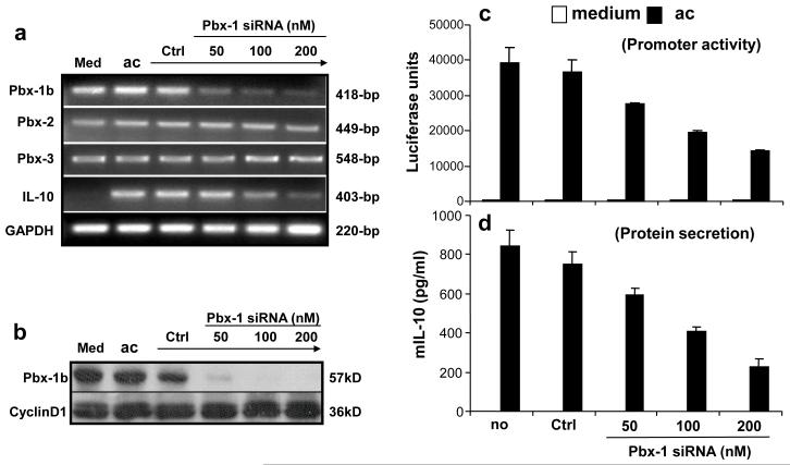
Figure 6. Blocking Pbx-1 expression inhibits apoptotic cell-induced IL-10 expression.
(a, b) Inhibition of Pbx-1 expression by siRNA. RAW264.7 cells were transfected by Oligofectamine with siRNA specific for Pbx-1 at various concentrations as indicated, or GFP (control) at 200 nM for 4 hrs, followed by exposure to apoptotic Jurkat cells. Eight hours later, total RNA was isolated and subject to RT-PCR for the analysis of Pbx-1a, Pbx-1b, Pbx-2, Pbx-3, IL-10, and GAPDH mRNA expression (a). Whole cell lysate were isolated and subject to Western blot analysis for Pbx-1b protein level (b) using a polyclonal antibody for Pbx-1, 2, 3.
(c, d) Inhibition of IL-10 expression by Pbx-1 siRNA. RAW264.7 cells were transfected with the IL-10 promoter construct by electroporation. Twelve hours later, the cells were transfected by Oligofectamine with Pbx-1 siRNA at various concentrations or the control siRNA at 200 nM for 4 hrs followed by exposure to apoptotic Jurkat cells. Luciferase activity (c) was measured 8 hr post exposure to apoptotic cells, and IL-10 protein secretion (d) was analyzed at 24 hr post stimulation.

