
Figure 4.
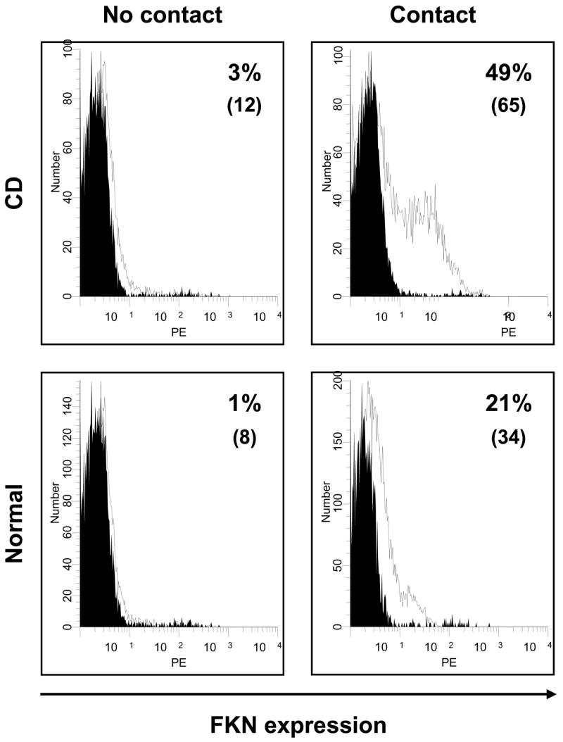
Leukocyte contact-induced upregulation of FKN surface expression by HIMEC. HIMEC monolayers were cultured with Jurkat T-cells for 24 hours, after which the percentage of FKN-expressing HIMEC was assessed by flow cytometric analysis. In some experiments (No contact), a 0.2 μm porous filter was placed between leukocytes and endothelial cells to prevent physical contact. Numbers in parentheses indicate the corresponding MFI. The black curve represents the background signal from the isotype control. This figure is representative of 12 separate experiments (4 normal, 4 UC and 4 CD HIMEC).