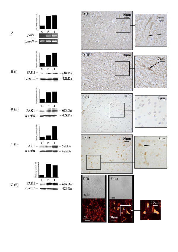
Figure 4.
PAK1 expression in human and rat brain following stroke. RT-PCR demonstrated an increase in PAK1 mRNA in infarcted and peri-infarcted areas of pooled samples from patients surviving from 2 to 6 days following stroke (A). Western blotting demonstrated an increase in protein levels in infarcted and peri-infarcted areas of patients surviving for 3 (Bi) and 15 (Bii) days following stroke and in rats at 12 h (Ci) and 24 h (Cii) following MCAO. Weak neuronal (axonal) staining (arrow) observed in contralateral areas of a patient surviving for 15 days following stroke (Di). Strong PAK1 staining in neurons (arrow) and cells with the morphological appearance of glia from infarcted areas of a patient surviving for 3 days following stroke (Dii). No staining observed in contralateral areas of rat brain at 24 h following MCAO (Ei) while strong PAK1 staining was seen in neurons (arrow) and cells with the morphological appearance of glia from infarcted areas of rat brain 1 h following MCAO (Eii). Stronger PAK1 immunofluorescent staining was seen in HFN following OGD (Fii) compared to control (Fi) (C: Contralateral, P: Peri-infarct, I: Infarct).

