
Figure 4.
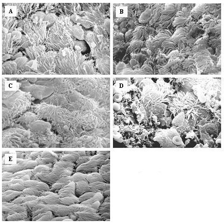
Scanning electron microscopy of the epithelial layer of the trachea from mice inoculated saline (E) or at various points after infection with C. pneumoniae: A: Day 2; B: Day 7; C: Day 14, D: Day 21 (2000–3000×). Two days after infection, the respiratory epithelium showed hyperthrophy of goblet cells and some scattered bacteria that were observed prevalently in contact with ciliated cells (Fig. 4A). After 7 days ciliary disorientation was the most evident change and there were a few single chlamydial bodies (Fig. 4B). The epithelium appeared severely damaged on day 14, goblet cells were notably hypertrophic and numerous bacteria were visible, also in little microcolonies (Fig. 4C). In addition to exfoliated epithelial cells on day 21 (Fig 4D), in some areas shorter cilia began to appear, a marker of ciliary regeneration.