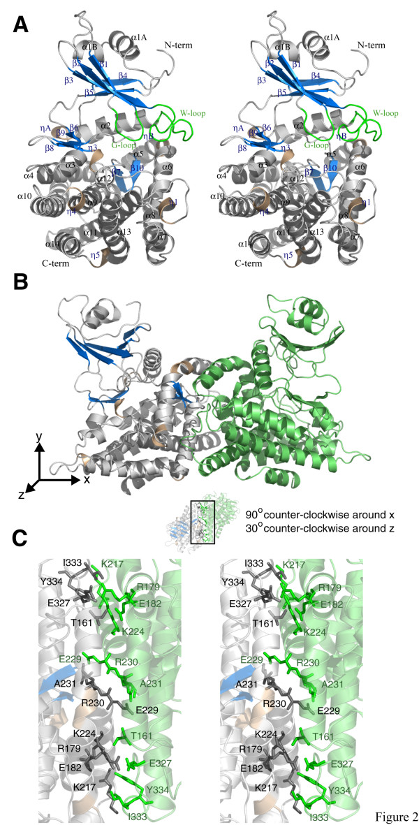
Figure 2.
Structure of A. thaliana MTR kinase. (a) Stereo representation of the plant MTR kinase monomer. Monomer A of the MTRK-ADP-MTR complex is shown, with the nucleotide and substrate omitted. α helices are represented as grey coils with the 310 helices as wheat coils. β strands are represented as blue arrows and loops as grey tubes. The G-loop and W-loop are coloured in green. (b) Dimeric structure of the complex with monomer A coloured as in (a) and monomer B in green. (c) Stereo representation of detailed interactions between the two monomers. Figure 2 was prepared using PyMOL [51].

