
Figure 1.
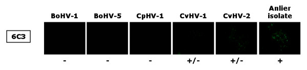
Indirect immunofluorescence staining of MDBK cells infected with either BoHV-1, BoHV-5, CpHV-1, CvHV-1, CvHV-2, or the Anlier isolate. Cells were incubated until viral plaques appeared and were then treated as described in Materials and Methods. 6C3 is the primary antibody and was detected by FITC-conjugated rabbit immunoglobulin anti-mouse IgG. Presence of viral plaques was checked by transmission microscopy before epifluorescence microscopy. All immunofluorescence stainings were performed three times. Symbols: +, positive signal; -, negative signal; +/-, weak signal.