
Figure 4.
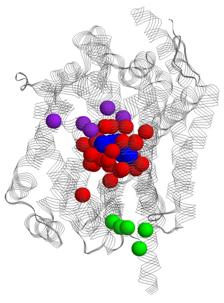
The entire LeuTAa structure is shown with the α-carbons of the 34 known functional sites highlighted; the binding site residues are colored red, cytoplasmic gate residues are colored green, and extracellular/periplasmic gate residues are colored purple. The leucine substrate and sodium ions are rendered in spacefill and colored blue.