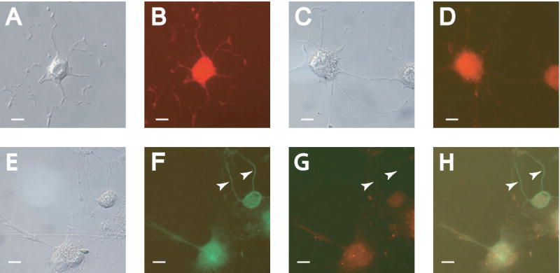
Figure 2. Staurosporine-induced differentiation leads to the development of dendrite-like neurites.

RGC-5 cells were differentiated for 48 hours and photographed with DIC optics (A, C, E), or under filters appropriate for immunocytochemical staining (B, D, F-H). (A-B) Differentiated cells showed staining for the neuronal marker β-III-tubulin strongly in all neurites. (C-D) Staining for the dendritic markers MAP2a and MAP2b revealed staining in the majority of neurites. (F). Labeling for all isoforms of MAP2 showed staining in all neurites. (G) Concurrent staining for the MAP2a and MAP2b isoforms revealed some unlabeled neurites. (H) Overlay of (F) and (G). Arrows indicate neurites showing exclusive labeling for MAP2c, suggesting an axonal phenotype. Scale bar in all panels indicates 10 μm.
