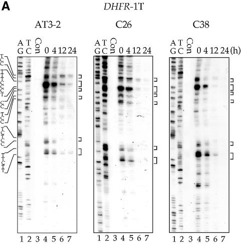
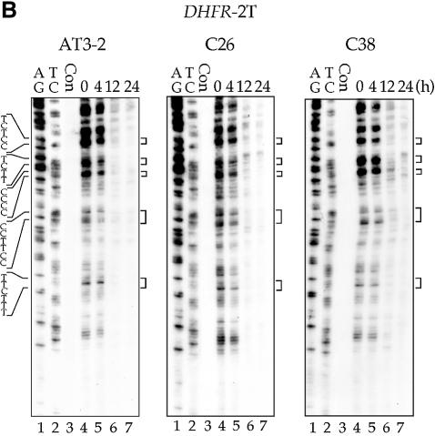
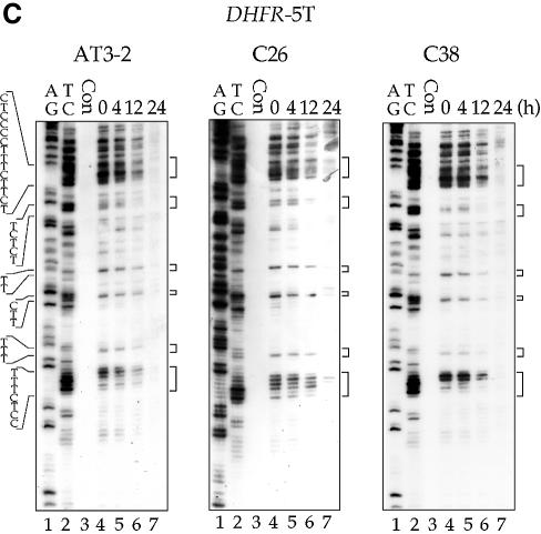
Figure 2.
The time course of CPD repair in the transcribed strand of exon 1 (A), exon 2 (B) and exon 5 (C) of the DHFR gene in CHO AT3-2, C26, and C38 cells. Cultured cells were UV irradiated (15 J/m2) and then incubated for various periods of time. Genomic DNA was isolated, treated with T4 endo V followed by photoreactivation, and then subjected to LMPCR. The LMPCR products were separated by electrophoresis in 8% denaturing polyacrylamide gels, transferred to nylon membranes, and hybridized with 32P-labeled probes specific for the transcribed strand of DHFR exons 1, 2 or 5. A + G and T + C represent Maxam–Gilbert sequencing reactions. Sequences of contiguous pyrimidines with the potential to form CPDs are indicated on the left, and T4 endo V incision sites are indicated on the right (bracketed). Lanes 4–7 show the relative frequency of T4 endo V cutting at dipyrimidine sites along each sequence at different post-UV repair time points (0, 4, 12 and 24 h). Lane 3 (Con) represents DNA isolated from unirradiated control cells and treated with T4 endo V. Very similar results were obtained from three independent experiments.


