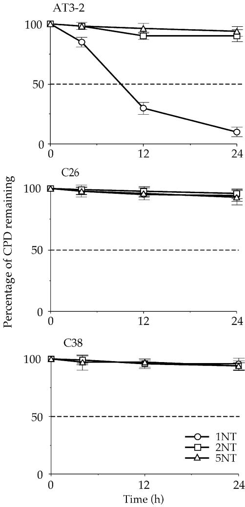
Figure 5.
The kinetics of CPD repair in the non-transcribed strand of the DHFR gene in AT3-2, C26 and C38 cells. The relative amount of CPD formed at the dipyrimidine sites (bracketed) along the non-transcribed strand of exons 1 (1NT), 2 (2NT) and 5 (5NT) of the DHFR gene for each time point shown in Figure 4 was quantified with a Cyclone Storage Phosphor System. The percentage of CPD remaining in the non-transcribed strand of each exon was plotted as a function of repair time. The results represent three independent experiments.

