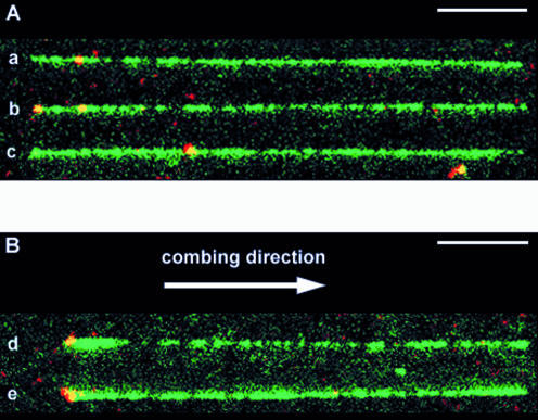
Figure 4.
Visualisation of labels on combed lambda molecules. DNA was stained with YOYO-1 (green). The labeled DNA fragments contained Alexa Fluor 546 (red); they therefore appear as yellow spots. The bars represent 5 µm. A montage of different patterns of combing and labeling is produced: (A) aligned longer (26–27.5 µm) molecules. The combed molecules were aligned so that the red internal labels were located on the left side, but they were equally distributed on both sides with respect to the combing direction (see text). (B) Aligned shorter molecules (23.5–25.5 µm). In this case, combed molecules were oriented with respect to the combing direction, i.e. the left side is the side where the DNA molecule sticks first to the glass surface, before being stretched in the direction indicated by the arrow.

