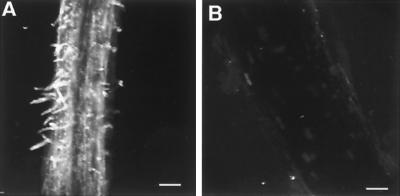
Figure 4.
Localization of LNP on D. biflorus roots. Immunofluorescence confocal microscopy of fixed whole mounts of the nodulation zone of 7-day-old D. biflorus roots treated with antiserum prepared against recombinant LNP (A) or preimmunization serum (B). Each image is a three-dimensional composite of 26–29 optical sections. (Bars = 100 μm.)

