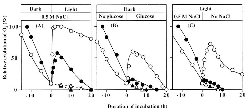
Figure 3.
Effects of light, glucose, and removal of NaCl on restoration of oxygen-evolving activity in wild-type and desA−/desD− cells after NaCl-induced inactivation. (A) Wild-type and desA−/desD− cells were incubated for 25 h and 12 h, respectively, in darkness in the presence of 0.5 M NaCl. Then cells were exposed to light of 50 μE⋅m−2⋅s−1 in the presence of 25 μg/ml lincomycin (broken lines) or its absence (solid lines). (B) Wild-type and desA−/desD− cells were incubated in darkness in the presence of 0.5 M NaCl as in A. Then glucose was added to a final concentration of 5 mM. (C) Wild-type and desA−/desD− cells were incubated with 0.5 M NaCl in light at 50 μE⋅m−2⋅s−1 for 45 h and 25 h, respectively. Then cells were collected by centrifugation, resuspended in fresh BG-11 medium with no added NaCl, and incubated in light. ○, Wild-type cells; ●, desA−/desD− cells in the absence of lincomycin; ▵, wild-type cells; and ▴, desA−/desD− cells in the presence of lincomycin. Each point represents the average of results from four independent experiments.

