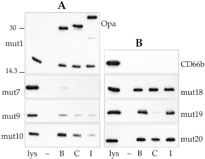
Figure 2.
Binding of His-tagged mutant CD66 N-domains to MS11 Opa variants. Shown are representative immunoblots of lysates derived from bacteria that had been incubated with His-tagged CD66 N-domain mutants. The upper blot in A was probed with anti-His and anti-Opa antibody; other blots with anti-His antibody only. Position of Opa proteins is indicated on the right (Opa). Molecular mass standards (in kD) are indicated on the left of the top panel in A. Lanes labeled lys indicate E. coli lysate, containing the appropriate N-domain in an amount that would be seen on the blot if 100% of the N-domain present in the lysate was bound by the gonococci. Lanes labeled –, B, C, and I indicate amount of N-domain bound by Opa−, OpaB-, OpaC-, or OpaI-expressing gonococci, respectively.

