
Figure 3.
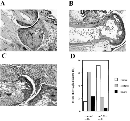
Histopathological assessment of arthritic joints. Mice treated with mGAL-1–transfected fibroblasts (4 × 10⁶ cells/mouse) showed mainly normal joints (A) and some moderately arthritic joints (B). Control mice treated with pCDNA3-transfected cells (4 × 10⁶ cells/mouse) showed moderate (B) and severe arthritis (C) accompanied by synovitis, erosions, and loss of joint integrity. A–C, hematoxylin and eosin staining; original magnification: ×100. Percentage of histological score per group is shown in D.