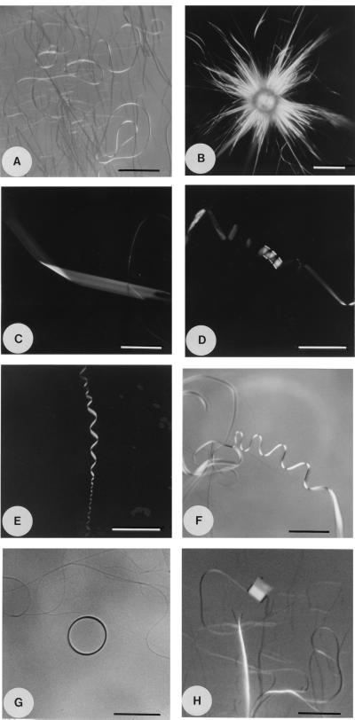
Figure 1.
Optical microscopy images (A–F and H: polarized light) of structures observed in a saturated 2,4,6-trichlorophenol-water solution. The solutions were prepared by heating an excess quantity of 2,4,6-trichlorophenol in water to about 80°C, followed by vigorous shaking and cooling to room temperature. The snapshots were taken at room temperature. (A) Filaments obtained in a vial and transferred onto the microscope slide (scale bar, 40 μm). (B–H) Structures obtained on the microscope slide in a drop of the initially isotropic solution. (B) Sheets and filaments forming on the edges of a disk-shaped, anisotropic phase (scale bar, 20 μm). (C) Winding of a ribbon-shaped sheet (scale bar, 40 μm). (D) A ribbon writhed into a three-turn, low-pitch helix (scale bar, 30 μm). (E) A multiturn, high-pitch helix (scale bar, 30 μm). (F) A ribbon twisting into a filament (scale bar, 40 μm). (G) A filament coiling into a tubule, viewed along its long axis (scale bar, 30 μm). (H) A side view of a nascent tubule (scale bar, 20 μm).

