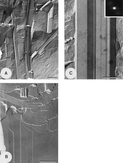
Figure 3.
Freeze-fracture electron microscopy of the filaments. A drop of the sample, about 20–50 μm thick, was deposited onto a thin copper holder, and the holder was either directly quenched in liquid propane, or the sample was first squeezed between the holder and a thin copper plate and then quenched in liquid propane. Both types of preparations were fractured in vacuo (about 10−7 torr) with the liquid nitrogen-cooled knife inside a Balzers 301 freeze-etching unit. The squeezed preparations were fractured by removing the upper plate with the cold knife. The replication was carried on by using the unidirectional shadowing with platinum carbon at an angle of 35°. The mean thickness of the metal deposit was 1–1.5 nm. The replicas were washed with ethanol and distilled water and then observed in a Philips EM 410 electron microscope. The contrast of the images depended on the fluctuations in the depth of the metal deposit. (A) Low magnification freeze-fracture electron micrograph of 2,4,6-trichlorophenol/water assemblies. Note the presence of very long filaments displaying well-defined longitudinal fracture planes (scale bar, 1 μm). (B) Higher magnification image of freeze-fractured 2,4,6-trichlorophenol/water filament showing periodically ordered, smooth lamellae (scale bar, 0.5 μm). (C) Part of a long filament fractured perpendicularly to the stacked lamellae. The optical diffraction inset shows the interlamellar spacing of 50 Å (scale bar, 0.25 μm).

