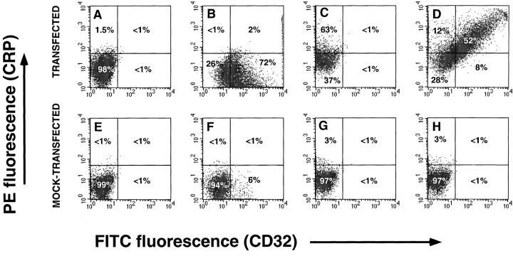
Figure 2.
Analysis of CRP binding to transfected COS-7 cells by two-color flow cytometry. COS-7 cells were transfected with the FcγRIIA plasmid, pcDSRα296. Mock-transfected cells received no DNA. Cells were incubated with 200 μg/ml of CRP and binding was detected with 2C10 and PE-GAM as described in Materials and Methods. Levels of FcγRII were determined by binding of FITC-AT10. A, B, C, and D, transfected; E, F, G, and H, mock-transfected. A and E, staining with 2C10 and PE-GAM; B and F, staining for CD-32 only; C and G, staining for CRP only; D and H, staining for CRP and CD32.

