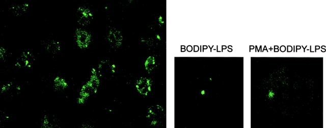
Figure 2.
Intracellular distribution of LPS in neutrophils. Optical sections of fluorescently labeled neutrophils are shown. PMN were incubated with both PMA (10 ng/ml) and BODIPY–LPS–sCD14 complexes (left and right) or BODIPY–LPS–sCD14 complexes only (center) for 30 min at 37°C. Comparing magnified cells of optical sections of neutrophils in the presence or absence of PMA shows that vesicular LPS labeling was not affected by the released specific granules, although the autofluorescence of PMN was increased with PMA stimulation.

