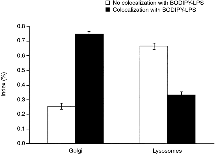
Figure 3.
Quantification of LPS with lysosomal and Golgi apparatus markers in neutrophils. Cells were labeled in the presence of either LysoTracker™ Red DND-99 or TRITC–CTB for 30 min at 37°C and processed for microscopy. 15–40 fields of live cells per experiment were observed, with an average of 5–20 cells per field. All green (LPS-containing) vesicles in the field were visually assigned as either not colocalized or colocalized with the red Golgi apparatus or lysosomal marker. Each experiment included data on 101–245 vesicles. Data is presented as the mean ± range for two experiments.

