
Figure 5.
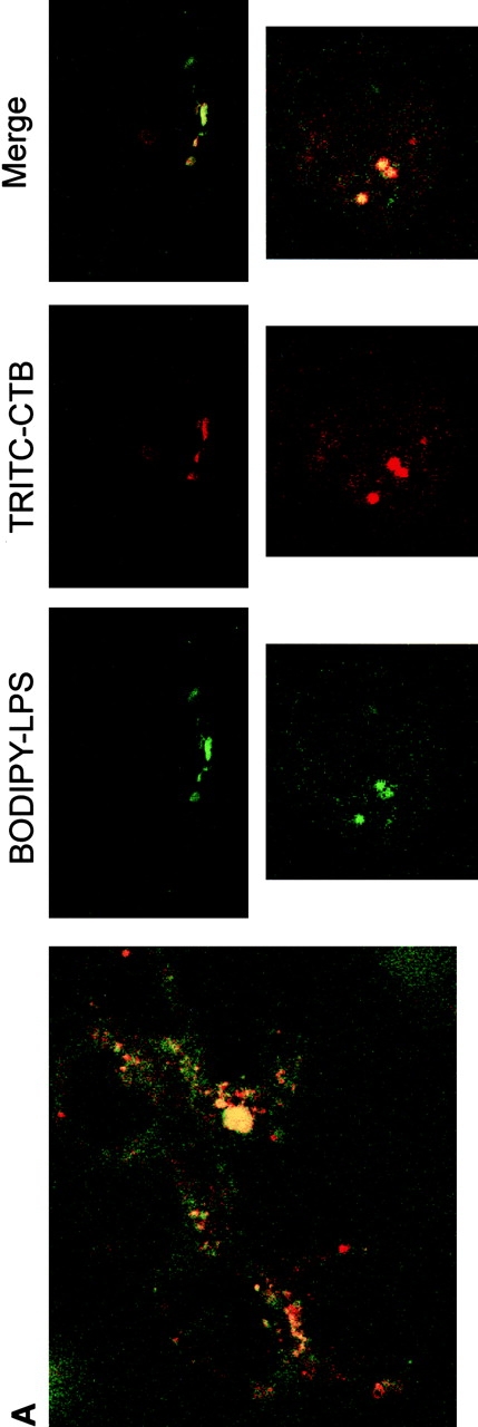
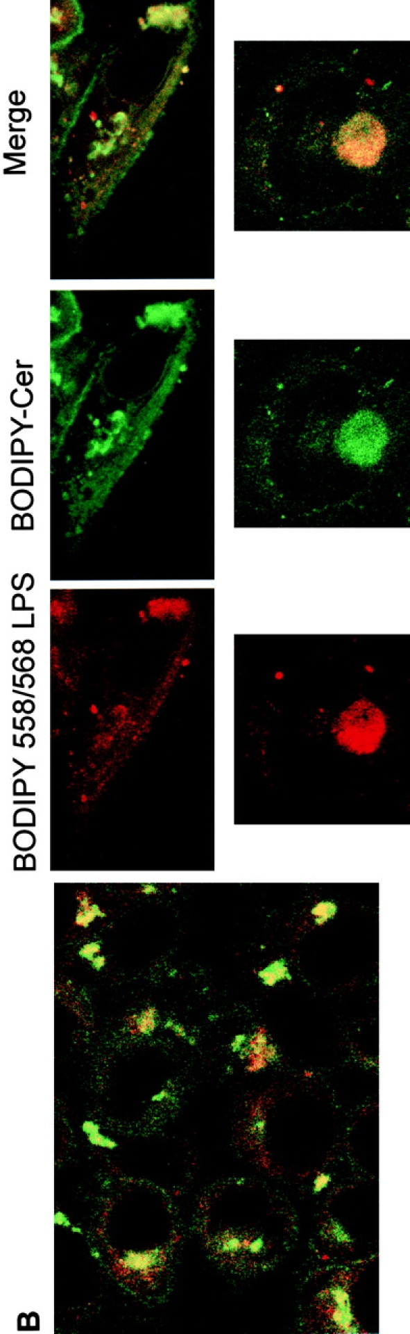
Intracellular distribution of LPS in HeLa cells. Optical sections of fluorescently labeled cells are shown. HeLa cells were incubated simultaneously with BODIPY–LPS and TRITC–CTB for 60 min at 37°C in DME with HSA and then processed for confocal microscopy. (A) LPS (green) is internalized and appears in a perinuclear area. Double-labeled HeLa cells reveal that BODIPY–LPS colocalizes with TRITC–CTB in the perinuclear area. (B) Cells were examined for staining of LPS (red) and ceramide (green). Merging the images revealed colocalization of LPS with ceramide (yellow), indicating that LPS colocalizes with Golgi apparatus markers.