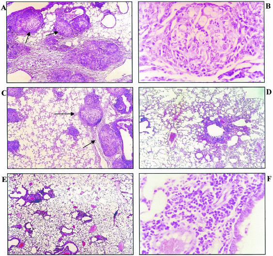
FIG. 10.
Histopathology of lung sections of P. brasiliensis-infected BALB/c mice 45 days after infection. (A) Control mice show well-organized granulomas (arrows) and a high number of yeast cells (magnification, ×100). (B) Granulomatous lesion from control group showing intact fungi and typical macrophages and epithelial cells (magnification, ×400). (C) Mice treated with MAb B7D6 show smaller, well-organized granulomas (arrows) with a high number of yeast cells (magnification, ×100). (D and E) Mice treated with MAb C5F11 (D) and mice treated concomitantly with both MAbs C5F11 and B7D6 (E) show absence of both yeast cells and granulomas (magnification, ×400). (F) Nongranulomatous lung from both MAb-treated mice, predominantly composed of a mononuclear infiltrate in which fungi are not seen (magnification, ×100).

