
FIG. 4.
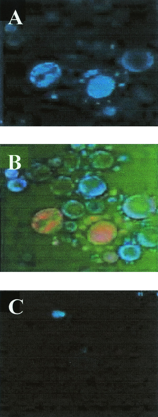
Confocal microscopy with anti-gp70 MAb revealed with mouse anti-IgG-fluorescein isothiocyanate (FITC). (A) gp70 labeling. (B) Double staining. Nuclei labeled with DAPI are blue, and gp70 is orange. (C) Negative control; absence of gp70 using only anti-mouse- FITC.