
Figure 2.

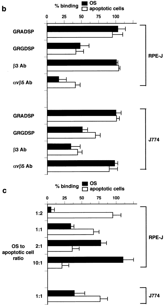
Apoptotic cells and OS compete for binding to αvβ3 of macrophages and for binding to αvβ5 of RPE cells. (a) Time course of combined OS binding plus internalization by rat bone marrow–derived macrophages (x) and NRK-F49 fibroblasts (•) compared with RPE-J cell combined binding plus internalization of OS (▾) and apoptotic cells (▿). J774 binding and internalization were indistinguishable from those of rat primary macrophages. (b) Integrin dependence and RGD peptide inhibition of binding of OS (black bars) and apoptotic cells (white bars) by RPE-J cells and by J774 macrophages. Mean OS binding indices in the absence of apoptotic cells were 6.3 ± 0.9 for RPE-J and 3.8 ± 0.4 for J774, after 2 h or 30 min of particle challenge, respectively. At these time points, particle phagocytosis accounted for <20% of total particle count. Mean apoptotic cell binding indices in the absence of OS were 5.5 ± 0.4 for RPE-J and 3.7 ± 0.5 for J774, at the same time points. (c) Quantitative competition of OS with apoptotic cells for phagocyte binding. RPE-J and J774 macrophages were challenged with different ratios of OS and apoptotic cells, for 2 h or 30 min, respectively. Particles were labeled with different fluorophores (FITC and Texas red) and quantified by fluorescence scanning. 100% represents binding of each particle in the absence of the other; for binding indices, see b. OS, black bars; apoptotic cells, white bars. Results represent means ± SEM (n = 5). When challenged with equal numbers of OS and apoptotic cells, both phagocytes seem to prefer apoptotic cells. However, this may be attributed to the larger size of apoptotic cells compared with OS (see Fig. 1).