
Figure 2.
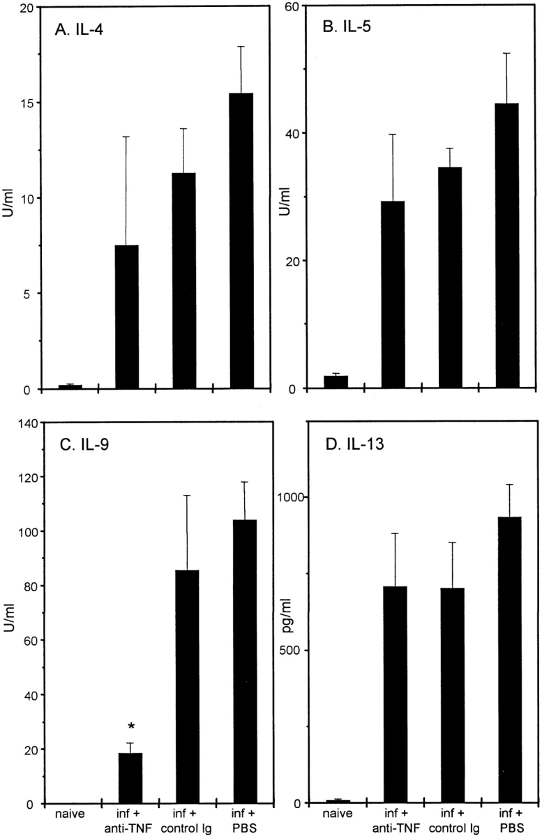
Cytokine production from T. muris–infected C57BL/6 mice after treatment with PBS, anti–TNF-α, or control Ig. Mesenteric lymph node cells were removed on day 21 p.i., stimulated in vitro with 50 μg T. muris Ag for 24 h, and supernatants were analyzed by sandwich ELISA for the presence of IL-4 (A), IL-5 (B), IL-9 (C), and IL-13 (D). Results represent the mean values of four mice per group ± SEM. *Significantly lower than control treated groups (P < 0.05).