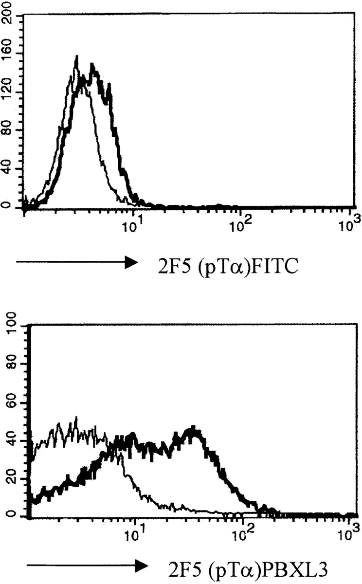
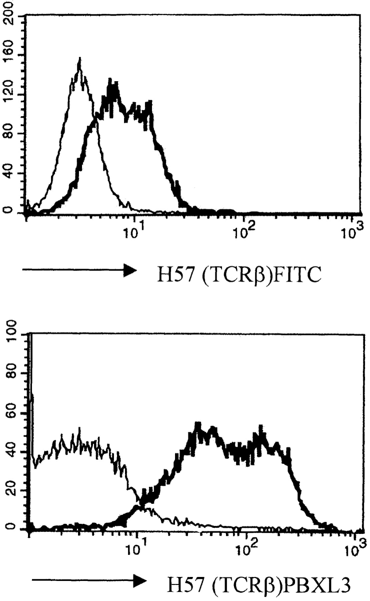
Figure 1.


Pre-TCR expression on the surface of the SCB29 cell line and comparison of the labeling intensity between streptavidin–PBXL-3 and streptavidin–FITC. SCB29 cells were initially incubated with biotinylated 2F5 (pTα) and H57 (pan-TCR-α/β) antibodies (thick line) or isotype-matched controls (thin line), and surface expression was revealed by streptavidin conjugated to the secondary reagents. Similar results were obtained in two other experiments.
